
Archived Content
The National Institute of Mental Health archives materials that are over 4 years old and no longer being updated. The content on this page is provided for historical reference purposes only and may not reflect current knowledge or information.
Brain’s Response to Scary Faces Imaged Faster Than You Can Say “Boo!”
• Science Update

Scientists have captured the split-second workings of the brain’s fear circuitry in people viewing frightful faces. NIMH researchers visualized this fleeting activity in the brain’s fear hub, called the amygdala, using a lightning-fast brain imaging technique called magnetoencephalography (MEG). They showed that such rapid, fear-related neural processes can now be studied non-invasively in living humans, with time resolution that other types of scanners can’t even come close to matching.
Background
Until now, scientists studying mental illnesses have been limited in their ability to see emotion circuits at work deep in the human brain. Brain circuits operate on a millisecond time-scale. Yet the predominant functional brain imaging tool, called functional magnetic resonance imaging (fMRI)*, can only see activity that lasts for at least one second. So fMRI studies could be missing some important action. By contrast, MEG tracks electro-magnetic activity, millisecond-by-millisecond. What remained to be demonstrated was whether MEG is sensitive enough to detect relatively weak signals emanating from deep seats of emotion – the amygdala and hippocampus.
To test MEG’s mettle, Brian Cornwell, Ph.D., Christian Grillon, Ph.D., of the NIMH Mood and Anxiety Disorders Program, and colleagues, scanned 14 healthy participants while they matched angry and fearful facial expressions, as well as geometric shapes, which served as a control. Previous fMRI studies had shown that viewing such faces triggers an amygdala response.
Results of This Study
Faces evoked greater activation of the left amygdala, in addition to other areas known to be involved in processing emotional faces. The non-emotional geometric shapes failed to similarly evoke this emotion-related circuitry. The scanner pinpointed the amygdala’s reaction with millisecond precision. A follow-up experiment with seven participants helped confirm that the response was traceable to the emotional content of the faces rather than to the identity of the actor portraying them.
Significance
Perfecting tools capable of capturing the human brain’s split-second response to a threatening stimulus has challenged neuroscientists. “Our results suggest that MEG can greatly enhance our ability to investigate rapid fear-related neural activity on a time scale impossible with other non-invasive brain imaging techniques,” said Cornwell.
What’s Next?
Another recent MEG study by NIMH researchers suggests that MEG may show promise as a tool for predicting antidepressant response. As they gain confidence in the technique’s ability to visualize emotional circuit activity, the researchers hope to make more use of it in studies of mood and anxiety disorders.
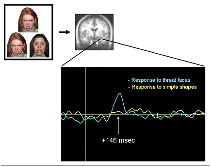
Participants in the MEG scanner matched angry and fearful facial expressions, which are known to activate the amygdala, and geometric shapes as a control. The MEG scanner registered an uptick in amygdala activity (blue blip) 146 milliseconds after a set of faces flashed on a screen (vertical white line). By contrast, a fMRI* scanner would only be able to detect a gradual rise in amygdala activity and the response would be delayed a few seconds. MEG holds promise for precise tracking of the brain’s emotional circuitry, the results suggest.
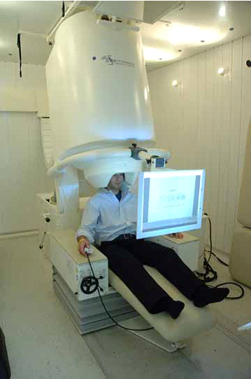
MEG scanner.
References:
Cornwell BR, Carver FW, Coppola R, Johnson L, Alvarez R, Grillon C. Evoked amygdala responses to negative faces revealed by adaptive MEG beamformers. Brain Res. 2008 Oct 7. [Epub ahead of print] PMID: 18930036
Cornwell BR, Johnson LL, Holroyd T, Carver FW, Grillon C. Human hippocampal and parahippocampal theta during goal-directed spatial navigation predicts performance on a virtual Morris water maze. J Neurosci. 2008 Jun 4;28(23):5983-90. PMID: 18524903
Tottenham, N., Tanaka, J., Leon, A.C., McCarry, T., Nurse, M., Hare, T.A., Marcus, D.J., Westerlund, A., Casey, B.J., Nelson, C.A. (in press). The NimStim set of facial expressions: judgments from untrained research participants. Psychiatry Research.
*Functional magnetic resonance imaging, fMRI, provides a picture of the brain's ever-changing activity rather than its static structure, as in anatomic MRI. This is accomplished by tracking the destination of oxygen carried in blood, which broadcasts a telltale radio signal generated by the magnetic field in the scanner. The more active a brain area is, the more oxygenated blood flows to it. The scanner picks up these signals and converts them into images. So fMRI provides an indirect, somewhat delayed picture of neural activity, with relatively high spatial resolution, but only moderate time resolution, compared to MEG.