
Archived Content
The National Institute of Mental Health archives materials that are over 4 years old and no longer being updated. The content on this page is provided for historical reference purposes only and may not reflect current knowledge or information.
Scans Reveal Faulty Brain Wiring Caused by Missing Genes
Grant Funds New Tools to Detect Subtler Wiring Errors in Schizophrenia
• Science Update
An NIMH study using an emerging imaging technology has discovered faulty wiring in the brains of people with Williams Syndrome, a rare genetic disorder that affects some aspects of thinking. Diffusion tensor imaging (DTI) scans revealed abnormal tracts of neuronal fibers that conduct long-distance communications between brain regions. The abnormalities likely resulted from neurons migrating to the wrong destinations during development, due to the absence of certain key genes, researchers say.
Such neuronal migrations gone awry are also suspected of playing a role in schizophrenia, although its genetic basis is less well understood than Williams Syndrome . To pinpoint the faulty wiring, NIMH-supported scientists at the University of Pennsylvania are perfecting an automated technique for analyzing DTI scans in schizophrenia, which is a more common mental disorder than Williams Syndrome, affecting one percent of adults.
DTI uses the same scanner hardware as conventional magnetic resonance imaging (MRI), but in a different way. Conventional MRI detects neuronal bodies, also called “grey matter,” to reveal brain anatomy. DTI detects neuronal extensions, also called “white matter,” to reveal the paths and directions of these nerve fiber tracts, which resemble bundles of thin pipes. White matter is so-named because, much like electrical wiring, the fiber tracts are protected by a white sheath of insulation. DTI detects the flow of water in the fibers, providing a measure of the structural connectivity between brain regions.
Karen Berman, M.D., Stefano Marenco, M.D., NIMH Genes Cognition and Psychosis Program, and colleagues reported on their DTI study of Williams Syndrome patients in the September 18, 2007, early online edition of the Proceedings of the National Academy of Sciences.
Williams Syndrome is caused by the deletion of some 28 genes in a particular section of chromosome 7. Among deficits characteristic of the syndrome are a lack of visual-spatial ability needed to assemble a puzzle, and a tendency to be overly-friendly with people, while overly anxious about non-social matters, such as spiders or heights. Many people with the disorder are also mentally challenged and learning disabled, but some have normal IQs.
The researchers built upon previous MRI and fMRI studies in which they identified brain regions that were both structurally and functionally abnormal in people with Williams Syndrome. They had reason to suspect that some of the deleted genes were important for development of fiber tracts. So they scanned five Williams Syndrome patients with normal IQs and compared them with matched healthy controls, using DTI.
As expected, the researchers found fiber tract abnormalities connected to the regions implicated in the earlier MRI/fMRI studies. These included excess fiber bundles, skewed fiber orientation and direction, and lopsidedness in the balance of white matter between the hemispheres (see graphic below).
The researchers say the findings provide new insight into how genes control development of white matter and can lead to normal or abnormal assemblies of neurons working together in the brain. They propose that certain genes deleted in Williams Syndrome code for proteins that would normally guide development and/or long-range connections of fiber tracks. The absence of these genes steers the tracts in the wrong direction, preventing them from reaching their natural targets in the brain during the later stages of neuronal migration – which results in the faulty wiring, suggest the researchers.
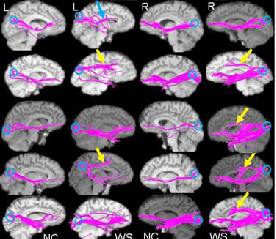
Diffusion tensor imaging scans show differences in brain wiring between Williams Syndrome patients (WS) and paired normal controls (NC). Four patients showed aberrant long-distance connections, or fiber tracts, coursing from the back of the brain to an area in the top front of the brain, particularly in the right hemisphere (yellow arrows). One patient had an aberrant tract projecting to a somewhat lower area in the mid front of the brain (blue arrow). Each row shows left and right views of long-distance fiber tracts in a patient and matched control set, as revealed by diffusion tensor MRI.
Source: Karen Berman, M.D., Stefano Marenco, M.D., NIMH Genes Cognition and Psychosis Program
High Math DTI Analysis Targets Schizophrenia
Compared to the gross fiber tract abnormalities of Williams Syndrome, discovering schizophrenia’s more elusive wiring errors tests the limits of existing DTI technology. With support from the NIMH Division of Neuroscience and Basic Behavioral Science, Ragini Verma, Ph.D., University of Pennsylvania, and colleagues, are developing an improved, automated system for analyzing DTI data from patients with schizophrenia, family members, and healthy controls. Their new method can detect several ways in which long-distance neural connectivity can be disrupted by disease. The goal is to gain a more complete picture of nerve fiber networks altered in schizophrenia than is possible with existing techniques. Obtaining these patterns of changes in the patients will help to characterize changes in their family members that may be related to the genetic roots of the disorder.
The Verma team’s mathematically sophisticated approach will leverage the statistical power gained from a large sample of 208 individuals to learn the underlying pattern of the nerve fiber tract abnormalities. Using more accurate and comprehensive measurements than are possible with conventional approaches, they hope to detect the subtle brain tissue abnormalities (predominantly white matter) likely to occur in schizophrenia.
Preliminary results from their ongoing study, based on scans of 34 patients with schizophrenia and 36 healthy controls, are revealing disruptions in connectivity and other abnormalities in networks responsible for attention and memory, voluntary muscle movements and motor skills, and regions that affect emotion processing. They have also observed gender differences relating to these abnormalities, according to Verma. Such differences could potentially help to explain sex differences in schizophrenia, such as why the age-of-onset is younger in males than females. The researchers also hope to correlate the abnormalities with symptom severity.
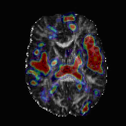
Composite diffusion tensor imaging scan image shows areas (red and to a lesser extent blue) where white matter fiber tracts and surrounding tissue differed between schizophrenia patients and controls.
Source: Ragini Verma, Ph.D., University of Pennsylvania, Department of Radiology-->
Marenco S, Siuta MA, Kippenhan JS, Grodofsky S, Chang WL, Kohn P, Mervis CB, Morris CA, Weinberger DR, Meyer-Lindenberg A, Pierpaoli C, Berman KF. Proc Natl Acad Sci U S A. 2007 Sep 18;104(38):15117-22. Epub 2007 Sep 7. PMID: 17827280
Parmeshwar Khurd, Sajjad Baloch, Ruben Gur, Christos Davatzikos, Ragini Verma. "Manifold Learning Techniques in Image Analysis of High-dimensional Diffusion Tensor Magnetic Resonance Images" Workshop on Component Analysis Methods for Classification, Clustering, Modeling, and Estimation Problems in Computer Vision, IEEE Conference on Computer Vision and Pattern Recognition (CVPR), June 2007.
- http://www.cs.cmu.edu/~ftorre/ca/program.html
- http://www.cs.cmu.edu/~ftorre/ca/ca_final_version/p18.pdf