
Archived Content
The National Institute of Mental Health archives materials that are over 4 years old and no longer being updated. The content on this page is provided for historical reference purposes only and may not reflect current knowledge or information.
$40 Million Awarded to Trace Human Brain’s Connections
Souped-up Scanners to Reveal Intricate Circuitry in High Resolution
• Press Release
The National Institutes of Health today awarded grants totaling $40 million to map the human brain's connections in high resolution. Better understanding of such connectivity promises improved diagnosis and treatment of brain disorders.
The grants are the first awarded under the Human Connectome Project . They will support two collaborating research consortia. The first will be led by researchers at Washington University, St. Louis, and the University of Minnesota, Twin Cities. The other will be led by investigators at Massachusetts General Hospital (MGH)/Harvard University, Boston, and the University of California Los Angeles (UCLA).
"We're planning a concerted attack on one of the great scientific challenges of the 21st. Century," explained Washington University's David Van Essen, Ph.D., who co-leads one of the groups with Minnesota's Kamil Ugurbil, Ph.D. "The Human Connectome Project will have transformative impact, paving the way toward a detailed understanding of how our brain circuitry changes as we age and how it differs in psychiatric and neurologic illness."
The Connectome projects are being funded by 16 components of NIH under its Blueprint for Neuroscience Research .
"On a scale never before attempted, this highly coordinated effort will use state-of-the-art imaging instruments, analysis tools and informatics technologies — and all of the resulting data will be freely shared with the research community," said Michael Huerta, Ph.D., of the National Institute of Mental Health, who directs the NIH Connectome initiative. "Individual variability in brain connections underlies the diversity of our thinking, perception and motor skills, so understanding these networks promises advances in brain health."
The Washington U./Minnesota team will map the connectomes in each of 1,200 healthy adults — twin pairs and their siblings from 300 families. The maps will show the anatomical and functional connections between parts of the brain for each individual, and will be related to behavioral test data. Comparing the connectomes and genetic data of genetically identical twins with fraternal twins will reveal the relative contributions of genes and environment in shaping brain circuitry and pinpoint relevant genetic variation. The maps will also shed light on how brain networks are organized.
In tooling up for the screening, the researchers will optimize magnetic resonance imaging (MRI) scanners to capture the brain's anatomical wiring - and its activity, both when participants are at rest and when challenged by tasks. All participants will undergo such structural and functional scans at Washington University. For these, researchers will use a customized MRI scanner with a magnetic field of 3 Tesla. This Connectome Scanner will incorporate new imaging approaches developed by consortium scientists at Minnesota and Advanced MRI Technologies and will provide ten-fold faster imaging times and better spatial resolution.
Additionally, a subset of twin pairs will also be scanned using more powerful 7 and 10.5 Tesla MRI units at the University of Minnesota, which has pioneered the use of such advanced, ultra high magnetic field imaging. For another subset of twins, the scans will be complemented by movies of millisecond brain electrical activity obtained at St. Louis University, using magnetoencephalography (MEG) and electroencephalography (EEG) .
After processing with sophisticated analysis tools using a supercomputer, the data will become web accessible via a customized Connectome Database Neuroinformatics Platform. All-told, the $30 million five-year project will involve 33 collaborators from nine research centers, including Oxford University, U.K.; Indiana University, Bloomington; University of California, Berkeley; Warwick University, U.K.; University d'Annunzio, Italy; and the Ernst Strungmann Institute, Germany.
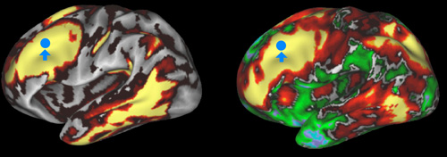
The Washington U. / U. Minnesota Connectome project will map the brain connections in each of 1200 participants. In the left image, yellow and red show a map of 'structural connectivity' in human cerebral cortex (regions that are connected to the blue spot as revealed by diffusion MRI). In the right image, yellow and red show a map of 'functional connectivity' (regions associated with the blue spot as revealed by functional MRI).
Also collaborating with this larger project, the MGH/Harvard-UCLA Connectome consortium will focus on optimizing MRI technology for imaging the brain's structural connections using diffusion MRI with unprecedented resolution. This way of using a MRI scanner, employed in both projects, maps the brain's fibrous long distance connections by tracking the motion of water. Different types of tissues are detectable by telltale water diffusion patterns characteristic of different types of cells. So the long extensions of neurons, called white matter, can been seen in sharp relief.
"The MRI scanner system we are assembling will be 4 to 8 times as powerful as conventional systems, enabling imaging of human neuroanatomy with much greater sensitivity than is currently possible," explained Bruce Rosen, M.D., Ph.D., who is co-directing the project with MGH/Harvard colleague Van Wedeen, M.D., and Arthur Toga, Ph.D., of UCLA.
The planned Connectome Scanner, to be built by Siemens Medical Systems for this project, is the first of a new class of MRI instruments. It will boost resolving power while also shortening the scan times required to image each subject, Rosen said.
The MGH/Harvard team has pioneered the use of a diffusion MRI technique called Diffusion Spectrum Imaging (DSI) to create stunning maps of neural fibers crisscrossing the brain. DSI offers a higher resolution, more multidimensional view than an older technique called Diffusion Tensor Imaging. This makes it possible, for example, to see the different orientations of multiple neural fibers that cross at a single location.
"Today we know less about the connectivity of the human brain than about a dozen other species," said Wedeen. "Learning more about variation in our own brain's connections will lay the groundwork for using brain imaging measures of connectivity as an aid in diagnosis."
"Creating these maps requires sophisticated statistical and visual informatics approaches," added UCLA's Toga. "Understanding the similarities and differences in these maps among sub-populations will improve our understanding of human brain in health and disease."
Supported by an $8.5 million grant over three years, the project will scan healthy adults, including some participants from the other consortia's project. Data and research know-how will also be shared across the two projects.
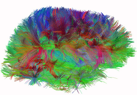
Diffusion spectrum image shows brain wiring in a healthy human adult. The thread-like structures are nerve bundles, each containing hundreds of thousands of nerve fibers.
About the National Institutes of Health (NIH): NIH, the nation's medical research agency, includes 27 Institutes and Centers and is a component of the U.S. Department of Health and Human Services. NIH is the primary federal agency conducting and supporting basic, clinical, and translational medical research, and is investigating the causes, treatments, and cures for both common and rare diseases. For more information about NIH and its programs, visit the NIH website .
NIH…Turning Discovery Into Health®
The NIH Blueprint for Neuroscience Research is a cooperative effort among the NIH Office of the Director and the 15 NIH Institutes and Centers that support research on the nervous system. By pooling resources and expertise, the Blueprint supports transformative neuroscience research, and the development of new tools, training opportunities, and other resources to assist neuroscientists.