Archived Content
The National Institute of Mental Health archives materials that are over 4 years old and no longer being updated. The content on this page is provided for historical reference purposes only and may not reflect current knowledge or information.
Magnetic Stimulation Boosts Human Memory, Network Connectivity
First Direct Evidence – NIH-supported Study
• Science Update
Scientists have improved memory for associations between faces and words by electromagnetically stimulating neural connections in a brain network. In the process, they produced the first direct evidence that stronger interactions between different areas of the human brain’s memory and thinking hubs underlie such associative memory.
“Our demonstration of a non-invasive method to target the function of deep brain structures could potentially be modified to treat disorders of hippocampus - cortex network connectivity, such as memory deficits in schizophrenia,” said Joel Voss, Ph.D., of Northwestern University, whose research was funded, in part, by NIMH. “However, substantial research will be required to move this technique from proof-of-concept to practical treatments.”
Voss, and colleagues, report on their functional neuroimaging study in the journal Science, August 29, 2014.
Evidence from animal research had suggested that associative memories are formed through interactions between the memory hub, the hippocampus, located deep in the brain, and a network of thinking circuitry in its outer mantle, or cortex.
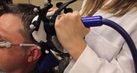
For each participant, Voss’s team first pinpointed the optimal site to stimulate over a brain area near the skull at the left side of the brain. They based this on the site’s connectivity with a target area deep in the memory hub during a resting state functional magnetic resonance imaging (fMRI) scan; fMRI provides a picture of which brain areas are active and connected at any given moment – the more in-synch the activity between sites, the more connectivity. Over the 5 days, the participants also performed a task in which they had to remember words associated with particular faces.
Both task performance and circuit connectivity improved after a few days of rTMS, but not after a similar course of sham, or placebo, rTMS. This confirmed that the stimulation did, indeed, account for the changes. The greater the connectivity among the network of brain areas, the more the subjects’ memory improved. The rTMS-triggered memory improvement lasted at least 24 hours.
The study confirms in human brain that memory depends on cortex areas working in concert with the hippocampus. It also demonstrates – for the first time – a way to non-invasively and specifically improve memory. Future trials will test the technique in people with memory impairments, said Voss.
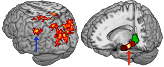
Cortex regions that showed increased connectivity with a part of the brain’s memory hub (left), or hippocampus (red arrow), following magnetic stimulation at sites over the left parietal cortex (blue arrow). Researchers linked improved performance on an associative memory task to the boost in connectivity. Source: Joel Voss, Ph.D., Northwestern University
Reference
Wang JX, Rogers LM, Gross EZ, Ryals AJ, Dokucu ME, Brandstatt KL, Hermiller MS, Voss JL. Targeted Enhancement of cortical-hippocampal brain networks and associative memory. Science, August 29, 2014. Vol. 345 no. 6200 pp. 1054 1057 DOI: 10.1126/science.1252900
See NIMH Director’s Blog: “Manipulating Memory”.
Grants
MH094263, NS083340