
Archived Content
The National Institute of Mental Health archives materials that are over 4 years old and no longer being updated. The content on this page is provided for historical reference purposes only and may not reflect current knowledge or information.
Schizophrenia, Autism Risk Gene Trajectories Point to Shared Causes
Monkey brain developmental atlas pinpoints when, where genes activate
• Science Update
Risk genes for schizophrenia and autism conspicuously activate in the same neuronal neighborhood of the brain’s cortex, or outer mantel, during infancy. This suggests some related underlying illness processes – even though known genetic variations associated with the disorders overlap by only 5 percent, say researchers.
Their study, which pinpointed the developmental trajectories of the suspect genes in the monkey brain, also identified divergent timing of risk gene activation that might help to explain the differing courses of the illnesses. Autism-related genes first switched on in newborn neurons during prenatal development, while schizophrenia risk genes didn’t activate until infancy.
This suggests that genetic risk for autism may have more to do with prenatal processes, such as the birth of new neurons, while schizophrenia risk genes may impact processes underway during infancy, such as refinement of neuronal circuitry. The delay is consistent with schizophrenia’s much later onset of symptoms in late adolescence/early adulthood, the researchers note.
The insights into the transcriptional origins of human mental illness were made possible by an NIMH-funded map of gene expression in the rhesus monkey brain across pre- and post-natal development. The map was developed by a scientific team of 100 researchers led by Ed Lein, Ph.D. , of the Allen Institute for Brain Science, who reported on their findings July 13, 2016 in the journal Nature.
The study focused on brain regions and developmental milestones linked to human mental illness. It turns out that only 9 percent of genes differ in their developmental trajectories between rhesus monkeys and humans – compared to 22 percent differing between rats and humans. Even though uniquely human disease and its specific brain circuitry can’t be modeled in non-human primates, comparing our brains with those of more closely related evolutionary cousins enhances understanding of human-specific features of brain development, say the researchers.
“This tremendous resource is freely available to the research community and will guide important research into the causes of many neurodevelopmental disorders for years to come,” said Michelle Freund, Ph.D., of the NIMH Office of Technology Development and Coordination, which funded the project.
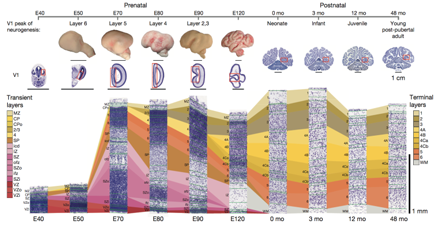
Primary visual cortex (outlined in red box, above) was analyzed for gene expression across 10 developmental stages from embryonic day 40 to 4 years after birth. Cortex layers are color coded and reflect the different sets of genes expressed across key developmental timepoints of the non-human primate brain. Source: Ed Lein, Ph.D., Allen Institute for Brain Research
A comprehensive transcriptional map of primate brain development.
Bakken TE, Miller JA, Ding SL, Sunkin SM, Smith KA, Ng L, Szafer A, Dalley RA, Royall JJ, Lemon T, Shapouri S, Aiona K, Arnold J, Bennett JL, Bertagnolli D, Bickley K, Boe A, Brouner K, Butler S, Byrnes E, Caldejon S, Carey A, Cate S, Chapin M, Chen J, Dee N, Desta T, Dolbeare TA, Dotson N, Ebbert A, Fulfs E, Gee G, Gilbert TL, Goldy J, Gourley L, Gregor B, Gu G, Hall J, Haradon Z, Haynor DR, Hejazinia N, Hoerder-Suabedissen A, Howard R, Jochim J, Kinnunen M, Kriedberg A, Kuan CL, Lau C, Lee CK, Lee F, Luong L, Mastan N, May R, Melchor J, Mosqueda N, Mott E, Ngo K, Nyhus J, Oldre A, Olson E, Parente J, Parker PD, Parry S, Pendergraft J, Potekhina L, Reding M, Riley ZL, Roberts T, Rogers B, Roll K, Rosen D, Sandman D, Sarreal M, Shapovalova N, Shi S, Sjoquist N, Sodt AJ, Townsend R, Velasquez L, Wagley U, Wakeman WB, White C, Bennett C, Wu J, Young R, Youngstrom BL, Wohnoutka P, Gibbs RA, Rogers J, Hohmann JG, Hawrylycz MJ, Hevner RF, Molnár Z, Phillips JW, Dang C, Jones AR, Amaral DG, Bernard A, Lein ES. Nature. 2016 Jul 13;535(7612):367-75. doi: 10.1038/nature18637. PMID:27409810