
Proton Spectroscopy of Rat Brain
For the in vivo studies using rat models, various localized proton spectroscopy techniques have been developed on an 11.7 T spectrometer with 89-mm vertical bore magnet. This high field spectrometer provides excellent signal-to-noise ratio and spectral resolution which allows user to observe certain NMR signals that are not detected in the lower field spectrometers.
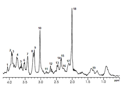
acquired using the adiabatic three dimensional
localization method (TR/TE = 5000/15 ms, 3.5 × 2.0 ×
4.5 mm3, NS = 128) from an oblique spectroscopy
voxel. The phosphocreatine methylene peak at 3.93
ppm and creatine methylene peak at 3.92 ppm are
clearly resolved. The labeled peaks are: 1
,5,6-myo-Inositol; 2-phosphocreatine; 3-creatine;
4, 17-glutamine+glutamate, 7,8-taurine;
9-choline-containing compounds; 1
0-phosphocreatine+creatine; 11-aspartate;
12,13,18-N-acetylaspartate; 14-glutamine;
15-glutamate; 16-gamma-aminobutyric acid;
19- lactate.