
Archived Content
The National Institute of Mental Health archives materials that are over 4 years old and no longer being updated. The content on this page is provided for historical reference purposes only and may not reflect current knowledge or information.
New Technique Pinpoints Crossroads of Depression in Rat Brain
Illuminates Brain Circuits at Work in Real Time
• Science Update
NIMH-funded scientists have developed a new high-speed technique for imaging brain activity and used it to pinpoint a circuit signal in rats that may be at the crossroads of depression — a possible "final common pathway" where different causes of, and treatments for, the disorder appear to converge. Activity in the circuit dampened when rats were in a depression-like state and revived after they were treated with antidepressants.
The technique, called voltage-sensitive dye imaging, reveals the split-second pace of brain circuits at work in real time and at the cellular level, as their electrical discharges make them glow.
"We detected circuit activity that accommodates disparate threads of evidence about depression," explained Karl Deisseroth, M.D., Ph.D., Stanford University. "This technique revealed a circuit signal where life experiences, biological vulnerabilities and treatments may come together — via diverse cellular mechanisms — to shape mood states."
Deisseroth, Raag Airan, Ph.D., Leslie Meltzer, Ph.D., and colleagues reported on their findings online July 5, 2007 in Science Express.
"This stunning demonstration of abnormal dynamics in a rat brain circuit required speed and sharpness far beyond techniques currently available for human studies," observed NIMH Director Thomas R. Insel, M.D. "We are not able to apply voltage-sensitive dye imaging to people with major depressive disorder, but studies that help us link behavior to real-time brain circuit information will be the foundation for understanding depression as a brain disorder."
Previous animal studies had found molecular and cellular markers for depression-like states, but not such a telltale circuit-level signal. Since the brain operates on a millisecond timescale and at a cellular level, the challenge was to overcome the coarse resolution and speed limitations of existing functional brain imaging techniques, most of which can't detect events briefer than a second or smaller than a millimeter. Voltage-sensitive dye imaging captures millisecond events with micrometer precision.
The Stanford team first created a depression-like state in rats, applying chronic mild stressors until the animals faltered in the face of challenging situations, much like the hopelessness experienced in depression.
The researchers then looked for any resultant circuit changes in the hippocampus, since previous studies had implicated this area in depression. The hippocampus regulates memory and mood. They soaked slices of hippocampus from depressed and normal rats in a fluorescent dye that glows in response the electrical discharges of brain cells. Then they electrically stimulated the slices and recorded movies of the circuit responses (see movie below). A smaller part of a hippocampus area called the dentate gyrus glowed in the slices from "depressed" rats than in slices from normal rats.
The glowing circuitry expanded to normal size in "depressed" rats that had been treated with an antidepressant (fluoxetine). This was accompanied by increased birth of new neurons in the dentate gyrus, adding to evidence from previous studies that such neurogenesis there is required for behavioral improvement following antidepressant treatment. Yet inhibiting neurogenesis failed to induce a depression-like state. So the dentate gyrus circuit signal revealed that even though neurogenesis could account for treatment response, it could not account for the depression-like state.
"This implied that depression and treatment can work via separate cellular mechanisms," explained Deisseroth. "But both funnel through a circuit-dynamics common pathway."
The dentate gyrus signal "may serve as a starting point in mapping the network-level changes in other brain regions implicated in depression," the researchers suggest.
Also participating in the research were: Madhuri Roy, Yuqing Gong and Han Chen, Stanford University.
Movie of circuit activity
Circuitry in rat brain dentate gyrus (part of the hippocampus) activates in response to pulses of electrical stimulation. The Stanford team found that the extent of such activation was diminished in rats conditioned to a depression-like state. They suggest that the dentate gyrus signal may be the brain's final common pathway for depression. They developed this technique, voltage sensitive dye imaging, to detect high-speed brain activity with micron resolution. Movie is slowed-down 17-fold from actual speed of circuit activity.
Source: Raag Airan, Stanford University
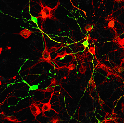
Deisseroth and colleagues identified brain circuit activity patterns that underlie depression-related behavioral changes. There was evidence that antidepressants may have normalized the behavior and circuit activity by triggering the birth of new neurons from adult neural stem cells (above; red = neurons; green = stem cell-derived cells, yellow = newborn neurons
Source: Leslie Meltzer, Stanford University
Reference
Airan RD, Meltzer LA, Roy M, Roy M, Gong Y, Chen H, Deisseroth K. High-Speed Imaging Reveals Neurophysiological Links to Behavior in an Animal Model of Depression. Science. 2007 Jul 5; [Epub ahead of print]