



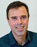
Peter Bandettini, Ph.D.
Research Topics
Functional MRI (fMRI) is a technique that utilizes the time-series collection of rapidly-obtained magnetic resonance images that are sensitive to brain activation-induced localized hemodynamic changes. Blood oxygenation level-dependent (BOLD) contrast is the most commonly used in fMRI because of its superior sensitivity and implementation efficiency.
The Section on Functional Imaging Methods (SFIM), in the Laboratory of Brain and Cognition (LBC), aims to enhance neuroimaging methods, and specifically MRI, by increasing the information content, functional precision, robustness, and interpretability of the acquired signals. Our group develops and tests novel approaches to fMRI acquisition, brain activation paradigms, processing methods, and multimodal integration. The overall goal is to better understand and utilize neuroimaging methods to increase our understanding of the human brain and to apply these methods and increased understanding to clinical diagnoses and treatments.
Our Section works along several related themes of development and inquiry. The first theme is that of developing and testing fMRI time series analysis using specific brain activation paradigms and physiological perturbations to derive novel information about subject’s cognitive state, task performance, brain physiology, cerebral spinal fluid fluctuations (including glymphatic system activity), as well as the similarities and differences between brain regions, subjects, or populations. The second theme is that of developing and testing novel MRI acquisition strategies and contrasts to enhance or quantify dynamic MRI signal changes during rest, activation, or physiologic manipulations. This second theme includes noise and artifact mitigation approaches including multi-echo fMRI. The third theme is to advance ultra high-resolution imaging at high field (7 Tesla) to better measure and utilize the information associated with cortical layer specific activity. We are engaged in creating and testing activation paradigms and hypotheses that would result in layer activity that is only measurable with layer-specific fMRI. Localizing brain activity at the cortical layer specificity allows the differentiation of feedforward activity from feedback activity, which provides unique insight to the brain’s hierarchal organization, thus enabling deeper insight into brain network organization. The fourth theme is to develop and curate neuroscience questions that most lend themselves to the cutting-edge acquisition and processing methods developed in our section and elsewhere in combination with multimodal measures including eye-tracking; heart and respiratory rate; pupillometry; peripheral measures of blood volume, oxygenation, pressure; behavioral measures; and simultaneous EEG or non-simultaneous EEG, optical imaging, and MEG. Attention to deriving information on similarities and differences between individuals – based on known traits is emphasized in all our themes.
SFIM consists of physicists, engineers, computer scientists, and neuroscientists who are either asking questions about healthy human brain function that push the limits of the technology or are advancing the technology in a way that may enhance our ability to ask deeper questions about the brain.
SFIM is unique in the NIH intramural program because it is focused both on developing methods and on understanding the functional organization and basic physiology of the human brain. We collaborate closely with the fMRI Core Facility, the Scientific and Statistical Computing Core, the Machine Learning team, the Data Sharing team, the Center for Multimodal Neuroimaging, as well as the Laboratory of Brain and Cognition, where the Section on Functional Imaging Methods resides.
Biography
Dr. Peter Bandettini received his Bachelor's degree in Physics in 1989 at Marquette University. He received his Ph.D. in Biophysics at the Medical College of Wisconsin (MCW) in 1994. His co-advisors were Drs. James Hyde and Scott Hinks. While in graduate school, he published two papers in Magnetic Resonance in Medicine. "Time course EPI of human brain function during task activation" was among the first to demonstrate fMRI, and "Processing strategies for time-course data sets in functional MRI of the human brain" introduced correlation analysis to fMRI and coined the acronym: FMRI.
From 1994 to 1996, Dr. Bandettini carried out his post-doctoral training under Drs. Jack Belliveau and Bruce Rosen at Massachusetts General Hospital and Harvard Medical School. After briefly returning to MCW as an Assistant Professor, he moved to Bethesda, MD in 1999 to work at the National Institute of Mental Health (NIMH) as a Principal Investigator and Director of the fMRI core facility. In 2016, he founded the Center for Multimodal Neuroimaging as well as the Machine Learning and Data Science and Sharing teams.
Dr. Bandettini has continuously worked to advance fMRI interpretability, precision, and utility. His research resides at the nexus of four fMRI domains: signal information content and interpretation, paradigm designs and processing methods, and finally, applications. He has advanced our understanding of the temporal, spatial, and interpretive opportunities and limits of fMRI and has helped foster the development of event-related fMRI, multi-contrast fMRI including multi-echo fMRI, multivariate analyses, dynamic resting state analysis, and cortical layer-resolved ultra-high resolution fMRI.
Dr. Bandettini has been fortunate to have highly gifted and motivated graduate students, post-docs, and staff scientists. He has fostered the talent in his lab through the encouragement of thinking in first principles, questioning assumptions, and confidently testing new ideas empirically. His trainees that have gone on to outstanding careers in academia include Drs. Natalia Petridou, Hauke Heekeren, Prantik Kundu, Rasmus Birn, Ziad Saad, Kevin Murphy, Niko Kriegeskorte, Laurentius Huber, Yuhui Chai, and Emily Finn.
Dr. Bandettini has been engaged in the leadership of both the MRI and Brian Mapping communities for over 30 years. He was president of the Organization for Human Brain Mapping (OHBM) from 2005 to 2006, and a member of the OHBM program committee for 14 years, which he chaired in 2002, 2011, and 2013. He also has served on the International Society for Magnetic Resonance in Medicine (ISMRM) program and education committees from 2007 to 2010, and young investigator award committee from 2001 to 2002. He was honored as ISMRM Fellow in 2015 and as OHBM Fellow in 2022.
In 2002, he received the OHBM Young Investigator Award. In 2020 he received the ISMRM Gold Medal, the Society's highest honor.
His 230+ papers and 20+ book chapters have been cited over 60k times. His h-index is 109 and has over 26 papers with more than 500 citations. He has co-edited a book on fMRI in 1999, and authored the book "fMRI," published in 2020. He has presented over 440 lectures worldwide.
He has been a leader in fostering scientific publishing and communication, having served as Editor-In-Chief of NeuroImage from 2011-2017. He is currently Editor-In-Chief of the OHBM Journal, Aperture Neuro, and since 2020, has been hosting a well-received OHBM-sponsored podcast: "Neurosalience."
Selected Publications
Huber L, Handwerker DA, Jangraw DC, Chen G, Hall A, Stuber C, Gonzalez-Castillo J, Ivanov D, Marrett S, Guidi M, Goense J, Poser BA, Bandettini PA (2017). High-Resolution CBV-fMRI Allows Mapping of Laminar Activity and Connectivity of Cortical Input and Output in Human M1. Neuron 96, 1253-1263.e7. https://doi.org/10.1016/j.neuron.2017.11.005. [Pubmed Link ]
Gonzalez-Castillo J, Saad ZS, Handwerker DA, Inati SJ, Brenowitz N, Bandettini PA (2012). Whole-brain, time-locked activation with simple tasks revealed using massive averaging and model-free analysis. Proc Natl Acad Sci U S A 109, 5487-92. https://doi.org/10.1073/pnas.1121049109. [Pubmed Link ]
Kriegeskorte N, Mur M, Bandettini P (2008). Representational similarity analysis - connecting the branches of systems neuroscience. Front Syst Neurosci 2, 4. https://doi.org/10.3389/neuro.06.004.2008. [Pubmed Link ]
Kundu P, Inati SJ, Evans JW, Luh WM, Bandettini PA (2012). Differentiating BOLD and non-BOLD signals in fMRI time series using multi-echo EPI. Neuroimage 60, 1759-70. https://doi.org/10.1016/j.neuroimage.2011.12.028. [Pubmed Link ]
Birn RM, Diamond JB, Smith MA, Bandettini PA (2006). Separating respiratory-variation-related fluctuations from neuronal-activity-related fluctuations in fMRI. Neuroimage 31, 1536-48. https://doi.org/10.1016/j.neuroimage.2006.02.048. [Pubmed Link ]
Magnuson Clinical Center, Room 1D80, MSC 1148
BETHESDA, MD 20814
Phone: +1 240 938 1610
Fax: +1 301 402 1370