Archived Content
The National Institute of Mental Health archives materials that are over 4 years old and no longer being updated. The content on this page is provided for historical reference purposes only and may not reflect current knowledge or information.
Genetic Tags Reveal Secrets of Memories’ Staying Power in Mice
• Press Release
A better understanding of how memory works is emerging from a newfound ability to link a learning experience in a mouse to consequent changes in the inner workings of its neurons. Researchers, supported in part by the National Institutes of Health’s National Institute of Mental Health (NIMH), have developed a way to pinpoint the specific cellular components that sustain a specific memory in genetically-engineered mice.
“Remarkably, this research demonstrates a way to untangle precisely which cells and connections are activated by a particular memory,” said NIMH Director Thomas Insel, M.D. “We are actually learning the molecular basis of learning and memory.”
For a memory to last long-term, the neural connections holding it need to be strengthened by incorporating new proteins triggered by the learning. Yet, it’s been a mystery how these new proteins – born deep inside a neuron – end up becoming part of the specific connections in far-off neuronal extensions that encode that memory.
By tracing the destinations of such migrating proteins, the researchers located the neural connections, called synapses, holding a specific fear memory. In the process, they discovered these synapses are distinguished by telltale molecular tags that enable them to capture the memory-sustaining proteins.
Mark Mayford, Ph.D., and Naoki Matsuo, Ph.D., of the Scripps Research Institute, report on their findings in the February 22, 2008 issue of the journal Science.
The Scripps researchers have been applying their new technique in a series of studies that focus on progressively finer details of the molecular machinery of memory.
“Inside neurons involved in a specific memory, we’re tracing molecules activated by that learning to see how it ultimately changes neural connections,” explained Mayford.
In a study published in the August 31, 2007 Science, Mayford and colleagues showed the same neurons activated by a learning experience are also activated when that memory is retrieved. The more neurons involved in the learning, the stronger the memory.
The researchers determined this by genetically engineering a strain of mice with traceable neurons in the brain’s fear center, called the amygdala. Inserted genes caused activated neurons to glow red when the animals learned to fear situations where they received shocks, in a process known as fear conditioning – and to glow green when the memory was later retrieved. The researchers then chemically prevented further expression of those neurons, so that resulting neural and behavioral changes could be confidently attributed to that learning experience at a later time. The study revealed which circuits and neurons were involved in the specific learning experience.
In the new study, Mayford and Matsuo adapted this approach to discover how fear learning works at a deeper level – inside neurons of the brain’s memory hub, called the hippocampus.
Evidence suggested that proteins called AMPA receptors strengthen memories by becoming part of the synapses encoding them. To identify these synapses, the researchers genetically engineered a strain of mice to express AMPA receptors traceable by a green glow. After fear conditioning had triggered new AMPA receptors deep in the neuron’s nucleus, they chemically suppressed any further expression of the proteins. This allowed time for the receptors to migrate to their appointed synapses. Hours later, green fluorescence revealed the fate of the specific AMPA receptors born in response to the learning.
As expected, the newly synthesized AMPA receptors had traveled and become part of only certain hippocampus synapses – presumably the ones holding the memory. Synaptic connections are made onto small nubs on the neuron called spines. These spines come in three different shapes called thin, stubby and mushroom. While little was known about the function of these differently shaped spines, the fact that they are altered in various forms of mental retardation, like Fragile-X syndrome, suggests a critical importance in mental function.
The researchers discovered the synapses that received the AMPA receptors with memory were limited to the mushroom type. The mushroom spines also figured prominently in the same neurons when the fear conditioning was reversed by repeatedly exposing the animals to the feared situation without getting shocked – a procedure called extinction learning. This indicated that the same neurons activated when a fear is learned are also activated when it is lost. The surge in mushroom spine capture of the receptors appeared within hours of learning and was gone after a few days, but appeared to be critical for cementing the memory.
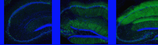
Newly synthesized AMPA glutamate receptors (green) were captured by mushroom-shaped spines in mouse hippocampus neurons encoding memory, after one (left), two (middle) and six (right) hours of fear conditioning. The receptors are thought to be key to strengthening the memory, making it long-term. The same green fluorescent protein that makes fireflies glow was genetically inserted into the receptors to reveal their destinations in the brain.
Source: Drs. Mark Mayford and Naoki Matsuo, Scripps Research Institute
Matsuo N, Reijmers L, Mayford M. Spine-Type Specific Recruitment of Newly Synthesized AMPA Receptors with Learning. Science. 2008 Feb 22;319(5866)
Reijmers LG, Perkins BL, Matsuo N, Mayford M. Abstract Localization of a stable neural correlate of associative memory. Science. 2007 Aug 31;317(5842):1230-3.PMID: 17761885
About the National Institute of Mental Health (NIMH): The mission of the NIMH is to transform the understanding and treatment of mental illnesses through basic and clinical research, paving the way for prevention, recovery and cure. For more information, visit the NIMH website.
About the National Institutes of Health (NIH): NIH, the nation's medical research agency, includes 27 Institutes and Centers and is a component of the U.S. Department of Health and Human Services. NIH is the primary federal agency conducting and supporting basic, clinical, and translational medical research, and is investigating the causes, treatments, and cures for both common and rare diseases. For more information about NIH and its programs, visit the NIH website .
NIH…Turning Discovery Into Health®