

Timing Diagram of Pulse Sequences for Proton Magnetic Resonance Spectroscopy at 7 Tesla
Glutamate is the main excitatory neurotransmitter in brain. Glutamine is synthesized from glutamate and ammonia. Both glutamate and glutamine participate in the glutamate-glutamine neurotransmitter cycle in which neuronal glutamate released into the synaptic cleft is taken up by astrocytes, converted to glutamine, and returned to neurons for replenishment of glutamate. Studies in humans and animals have found altered glutamate and glutamine levels in many brain disorders. Glutathione is an antioxidant and detoxifier. Glutathione was implicated in several neuropsychiatric diseases related to oxidative stress.
In proton magnetic resonance spectroscopy, the glutamate signal at 2.35 ppm, glutamine signal at 2.45 ppm, and the signal of the glutamyl moiety of glutathione at 2.54 ppm overlap each other at low magnetic fields. We have developed a 7 Tesla proton magnetic resonance spectroscopy technique to spectrally resolve glutamate, glutamine, and glutathione. At the same time, the interfering signal from the aspartyl moiety of N-acetyl-aspartate was eliminated using a novel J suppression scheme. Our method places a radiofrequency pulse at the resonance frequency of the N-acetyl-aspartate aspartyl methine proton at 4.38 ppm, thereby altering the J evolution of the N-acetyl-aspartate aspartyl methylene multiplet at 2.49 ppm. This method suppresses the 2.49 ppm signal in a single shot, allowing highly precise in vivo detection of glutamate, glutamine, and glutathione in the human brain.
Pulse sequence described in An L, Li S, Murdoch JB, Araneta MF, Johnson C, Shen J. Detection of glutamate, glutamine, and glutathione by radiofrequency suppression and echo time optimization at 7 tesla. Magnetic resonance in medicine 2015;73:451‐458.
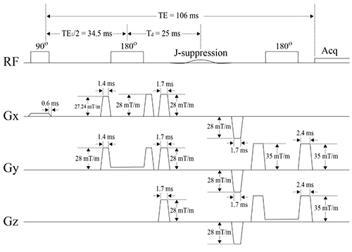
- Amplitude‐modulated excitation pulse; duration = 4.5 ms; B1max = 18.6 µT; FWHM = 3.1 kHz; rephase fraction = 0.1666.
- Amplitude‐modulated refocusing pulse; duration = 8.0 ms; B1max = 18.6 µT; FWHM = 2.0 kHz.
- Amplitude‐modulated J‐suppression pulse; frequency = 4.38 ppm; flip angle = 90° ; duration = 20 ms; B1max = 1.19 µT; FWHM = 158 Hz.
- Ramp‐up and ramp‐down times for all gradients are 0.6 ms.
- ADC: spectral width = 4000 Hz; number of data points = 2048; duration = 512 ms
- TR = 2.5 s.