Higher-Order Magnetic Field Shimming
Homogeneous magnetic field is a critical factor for spectral resolution and signal-to-noise ratio. Higher order shims are used when the inhomogeneous magnetic field is strong and is unable be compensated by the linear shims.
There is an existing high order shim method, FASTMAP, which samples the magnetic field along a group of radial columns which focus on the center of a localized voxel. It is a method for optimizing the field homogeneity in a cubical local region. However, is usually not implemented on clinical scanners. We have implemented this method on GE’s scanner and used it for 13C spectroscopy.
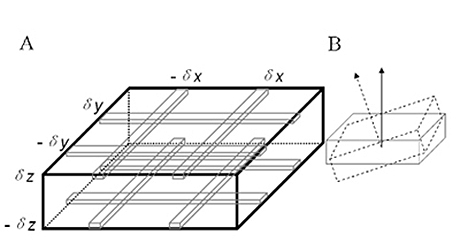
When the shim region is not a cubical space, for example a rectangular slab, the FASTMAP may not be the best shimming tool. We developed a variant of FASTMAP, using a group of parallel columns which forms a rectangular slab, see Fig. 12.

represented by x = dx, x = -dx , y = dy, y = -dy. Upper and
lower slices are denoted by z = dz and z = -dz, respectively.
(B): An oblique slice in physical frame r´ is in an axial plane in
logical frame r.
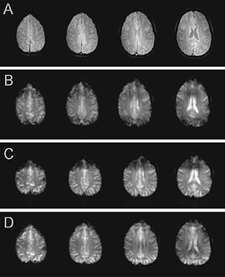
It was found that a pair of four columns in two separate slices could determine an optimal correction field comprising the spherical harmonic terms up to the third-order. The technique of multiple stimulated echoes was incorporated into the method, allowing the use of at least eight shots to accomplish field mapping. The shim currents were first determined in the logic frame by assuming that the slices were in axial planes, and then uniquely converted into the physical frame where the slices could be at any oblique angle, by using a spherical harmonics rotation transformation. This method thus works regardless of slice orientation. It was demonstrated on a 3 T scanner equipped with a complete set of second-order harmonic shim coils, see Fig. 13. Both phantom and in vivo experiments showed that this newly introduced high-order shimming method is an effective and efficient way to reduce field inhomogeneity for a region of imaging slices.

anatomic images (A), after manufacture’s first-order shimming
(B), after manufacture’s high-order shimming (C) and after
PACMAP (D). The EPI acquisition parameters were: single shot,
matrix size = 80×80, TE = 21.3 ms, slice thickness = 4.5 mm,
slice spacing = 2.5 mm and receiver bandwidth = 166 kHz.
Reference: Zhang Y, Li S, Shen J: "Automatic High-Order Shimming Using Parallel Columns Mapping (PACMAP)." Magn. Reson. Med. 62, 1073-1079 (2009).

