

Excellent to the “Core”: World Class Neuroimaging at NIMH
Peter Bandettini, Ph.D., Chief of the Section on Functional Imaging Methods and Director of the Functional Magnetic Resonance Imaging Core Facility, on behalf of the NIMH Intramural Research Program
• 75th Anniversary
For 75 years, NIMH has transformed the understanding and treatment of mental illnesses through basic and clinical research—bringing hope to millions of people. This Director’s Message, guest-written by NIMH’s Intramural Research Program, is part of an anniversary series celebrating this momentous milestone.
Our sensations, thoughts, and emotions are grounded in internal physical processes that are difficult to see and measure, much less understand. For well over a century, the links between what is happening in our brains and how we experience the world have started to emerge as brilliant innovations that allow more insightful questions.
As far back as the late 1800s, links between brain lesion locations and specific functional deficits were starting to be identified. Other means for measuring physiologic changes with brain activity were also being developed, ranging from precise balances that detected subtle increases in blood volume in the brain associated with increased mental activity to scientifically rigorous detection in animal models of blood volume changes, showing signal changes that were stunningly like functional magnetic resonance imaging (fMRI) signals now commonly seen 100 years later.
In the past 50 years, the National Institute of Mental Health (NIMH)'s Division of Intramural Research Programs (IRP) has been a world leader in developing and implementing advanced brain imaging technology. The division has fostered world-class facilities and seminal scientific advances that have allowed us to delve into the complex circuitry of the human brain. We’ve started shedding light on mechanisms and principles of brain function and are uncovering disease signatures and potential inroads for treatment.
Early innovations in brain imaging
A powerful imaging approach known as positron emission tomography (PET) is used today in research and clinical practice to create a map of metabolic processes or neurotransmitter activity in the brain. This technology was pioneered in the 1970s by NIMH IRP researcher Louis Sokoloff, M.D., Ph.D. He developed a method by which a radioactive tracer accumulated in specific brain regions in proportion to more active areas. He collaborated with computer scientists at NIMH to create a computer program that could measure this radioactivity and translate it into color-coded images representing brain activity. By the end of the 1970s, this technology had been adapted for use with humans, and PET scanners became available for use in medical and research settings.
The tools developed by Dr. Sokoloff have been refined over the years and are still in use today. For example, NIMH researcher Robert Innis, M.D., Ph.D., in collaboration with radiochemist Victor Pike, Ph.D., and colleagues are using a state-of-the-art PET facility at the National Institutes of Health (NIH) to develop a new way to deliver radioactive markers to brain cells, improving the mapping of neuroinflammation in the brain. This advance will allow researchers to better understand the role of neuroinflammation in a range of brain-based disorders from depression to Alzheimer’s disease.
The birth of core facilities at NIMH
Since the 1980s, NIH has been at the forefront of developing and nurturing resources to push the boundaries of brain science. Neuroimaging instrumentation can be expensive, and true expertise is rare. Having dedicated, shared groups, or “core facilities,” that develop sophisticated methods and collaborate with other scientists has proven to be an effective way to incubate breakthrough experiments and produce key findings.
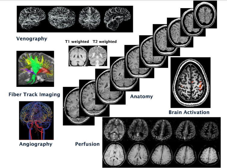
An early shared resource, the NIH-wide Nuclear Magnetic Resonance Center, was created in 1987. It was here that seminal early work pioneering MRI was performed. Robert Turner, Ph.D., helped pioneer blood-oxygen-level-dependent contrast used in the most common noninvasive brain imaging methods today. Peter Basser, Ph.D., along with Denis Le Bihan, M.D., Ph.D., invented diffusion tensor imaging , which was fundamental to the creation of the spectacular fiber track images used today to reveal the connectivity in the human brain.
Decades of discovery, collaboration, and growth
The NIMH IRP has led the way in creating and supporting core facilities, helping push the boundaries of neuroscience research.
One example is the proliferation of fMRI within NIH. The discovery of fMRI in the early 1990s revolutionized brain imaging and neuroscience. NIMH and the National Institute of Neurological Disorders and Stroke (NINDS) initiated an MRI facility to advance this exciting technology and to accommodate adventurous neuroscience research groups wanting to perform MRI and fMRI—peering into the living human brain not only to understand its structural details but now, thanks to fMRI, to observe functional activation with unprecedented speed, resolution, and fidelity. In 1999, Peter Bandettini, Ph.D., was recruited by NIMH to direct the new Functional MRI Facility (FMRIF) and lead his own research group in developing fMRI methodology, interpretation, and applications.
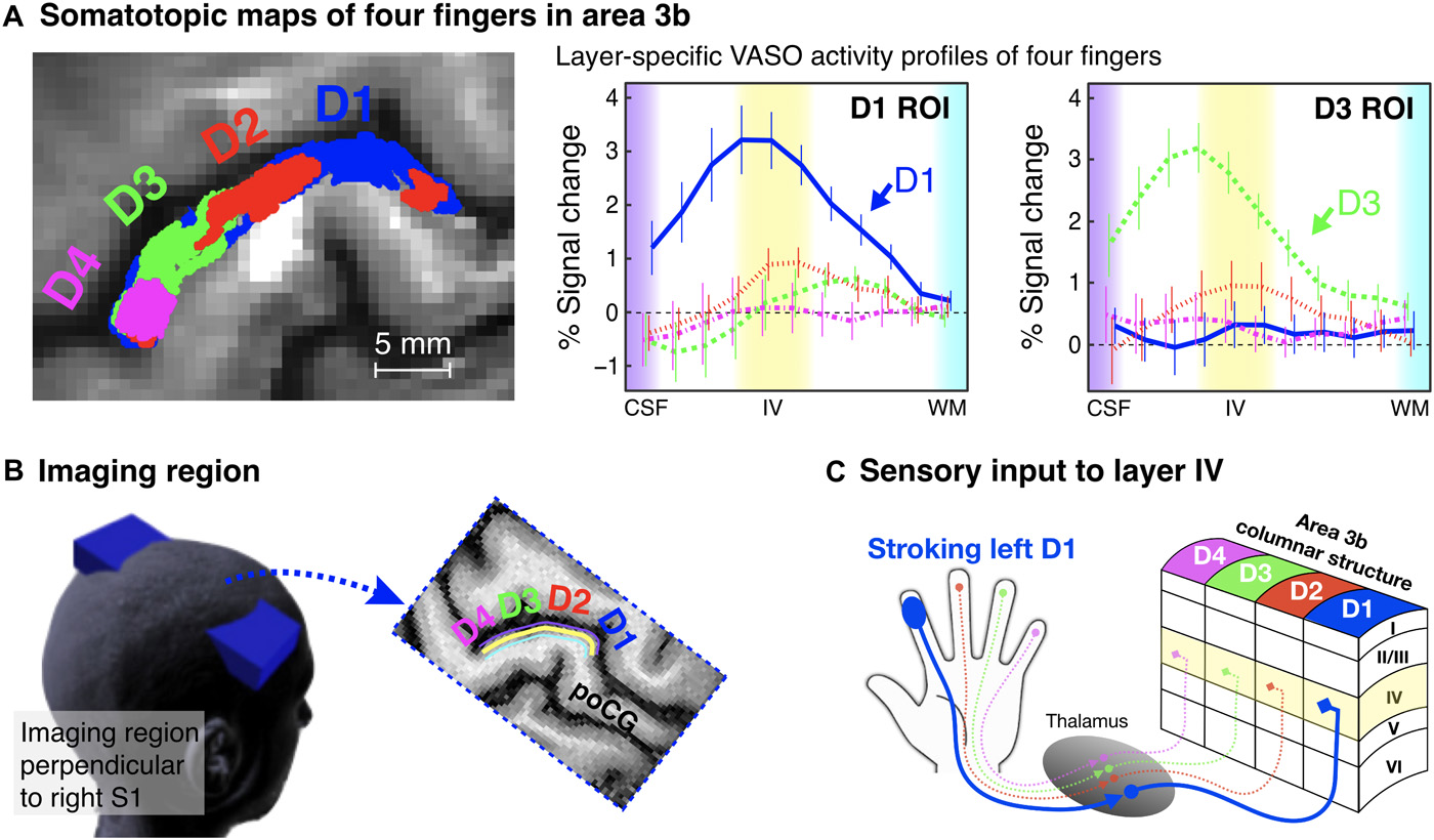
Over the years, the FMRIF has developed and adopted the latest capabilities and services for a rapidly growing number of investigators who want to use fMRI or MRI in their brain research. For example, the image to the right shows exquisite resolution fMRI of detailed cortical circuitry activation with individual finger activation. Not only is fMRI able to delineate the somatosensory activation with each finger, but it can show, from the peaks in the graphs, that the activation is in layer IV in the somatosensory cortex, which has only been known from research with animals to be selectively active with stimulation.
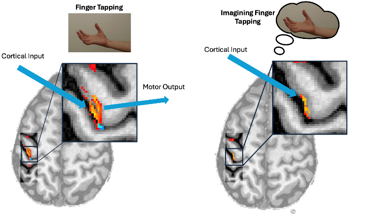
In another example, the image to the left shows two cortical layers becoming active for finger tapping and only one layer active for imagining of finger tapping. This higher resolution has tremendous potential to allow the creation of more detailed and informative maps of brain function—delving ever more into the complex circuitry and potentially revealing subtle differences associated with disorders.
Additional NIMH core facilities that develop and disseminate the latest neuroimaging technology to IRP scientists have rapidly emerged following the establishment of the fMRI core facility.
- In 2000, NIMH created a Neurophysiology Imaging Facility for nonhuman primate imaging, led by David Leopold, Ph.D.
- In 2001, Robert Cox, Ph.D., established the Scientific and Statistical Computing Core to help develop and maintain a brain imaging software platform called Analysis of Functional NeuroImages (AFNI), widely used by researchers to analyze MRI and fMRI data. The core is now led by Paul Taylor, Ph.D.
- Also in 2001, the Magnetoencephalography Core Facility was created to focus on research using magnetoencephalography and electroencephalography. This core facility was initially led by Rich Coppola, DSc., and is now led by Allison Nugent, Ph.D. Dr. Coppola was also instrumental in working with Dr. Sokoloff in the early days, converting his metabolism maps to color images.
- In 2002, the Magnetic Resonance Spectroscopy Core Facility, led by Jun Shen, Ph.D., was added to provide support for researchers using magnetic resonance spectroscopy, a method for identifying specific metabolite concentrations in the brain.
- In 2016, the Machine Learning Team, led by Francisco Pereira, Ph.D., and the Data Science and Sharing Team, led by Adam Thomas, Ph.D., began helping IRP investigators analyze and share their data more broadly.
- Research groups focusing on imaging methods, including the Noninvasive Neuromodulation Unit headed by Sarah Hollingsworth “Holly” Lisanby, M.D., and the Section on PET Neuroimaging Sciences led by Robert Innis, Ph.D., round out the NIMH’s vast neuroimaging resources. Neuromodulation is a method by which the brain is stimulated with high spatial and temporal accuracy—either for treatment or research.
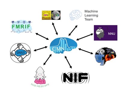
- In 2016, a Center for Multimodal Neuroimaging was created to link all the imaging-related cores together. This center serves as a nexus where the neuroimaging groups can come together to find points of fruitful intersection.
NIMH core facilities today
Today, the brain imaging core facilities remain at the forefront of science, helping NIH researchers undertake cutting-edge work. For example, the fMRI core facility has five MRI scanners, which are managed by five physicist staff scientists and used by more than 30 principal investigators performing both clinical and basic human neuroimaging research. Such a concentration of scanning technology and expertise cannot be found anywhere else in the world. Of note are the two 7T scanners in the core facility. These are the most powerful scanners approved for human use; only about 120 are available worldwide. Such powerful scanners enable NIH researchers to address entirely new questions about human functional brain organization, connectivity, and hierarchy and measure subtle differences and similarities between individuals.
Embraced by NIMH, the concept of core facilities and teams marked a paradigm shift for the institute. Cores scale with demand and foster expertise and strong collaborations with and between principal investigators. The resulting science and productivity demonstrate their success. Since 2000, NIH IRP investigators published more than 1,000 papers using the fMRI core facility. NIH and NIMH IRP investigators have just begun exploring the possible questions that may be asked using this unprecedented collection of world-leading tools.
NIMH has some of the most sophisticated tools for peering into the human brain. It is also home to world-class scientists, engineers, programmers, and clinicians who develop and wield these powerful instruments. The goal is to bridge that elusive connection between our physical brains and our sensations, thoughts, and emotions. The ultimate goal is that, with a deep and precise understanding of these connections, we can make more rapid headway toward improving mental health.